2026-02-25
Overview
In the last five years, the field of cardiac imaging has moved faster than perhaps any other medical specialty. Gone are the days when a simple 2D Echo or a diagnostic catheterization were the only tools in a cardiologist's arsenal. By 2026, the focus has shifted entirely from reacting to heart disease to predicting it. Non-invasive technologies like CCTA (Coronary CT Angiography) and AI-enhanced MRI have become the new gold standards.
But as scanner technology advances, a new challenge has emerged for hospitals and heart centers: Data Overload. Here is how cardiac imaging has evolved this year and why your software infrastructure (CVIS) matters more than your hardware.
How Cardiac Imaging Has Evolved in 2026
1. The Rise of One-Stop Non-Invasive Diagnostics
As noted in recent clinical studies, 2026 has seen a massive reduction in purely diagnostic catheterizations. Why invade the body when cardiac imaging can see everything from the outside?
FFR-CT Integration: We can now assess blood flow and blockages (Fractional Flow Reserve) purely through CT scans, without a wire.
4D Flow MRI: Magnetic Resonance Imaging now visualizes blood flow dynamics in real-time, offering insights into valvular disease that were impossible to see a decade ago.
The Software Challenge: These advanced scans generate gigabytes of data per patient. Legacy PACS systems crash under the weight of 4D datasets. You need a specialized Cardiovascular Information System (CVIS) like NeoRad that is built to handle high-frame-rate, dynamic imaging without lag.
2. AI Has Become the Doctor's Second Opinion
In 2026, AI in cardiac imaging is no longer a pilot program, but a must-have for the workflow. When a patient gets an Echocardiogram nowadays, it is assumed that the program will:
- Auto Contour: The software automatically draws the heart chambers.
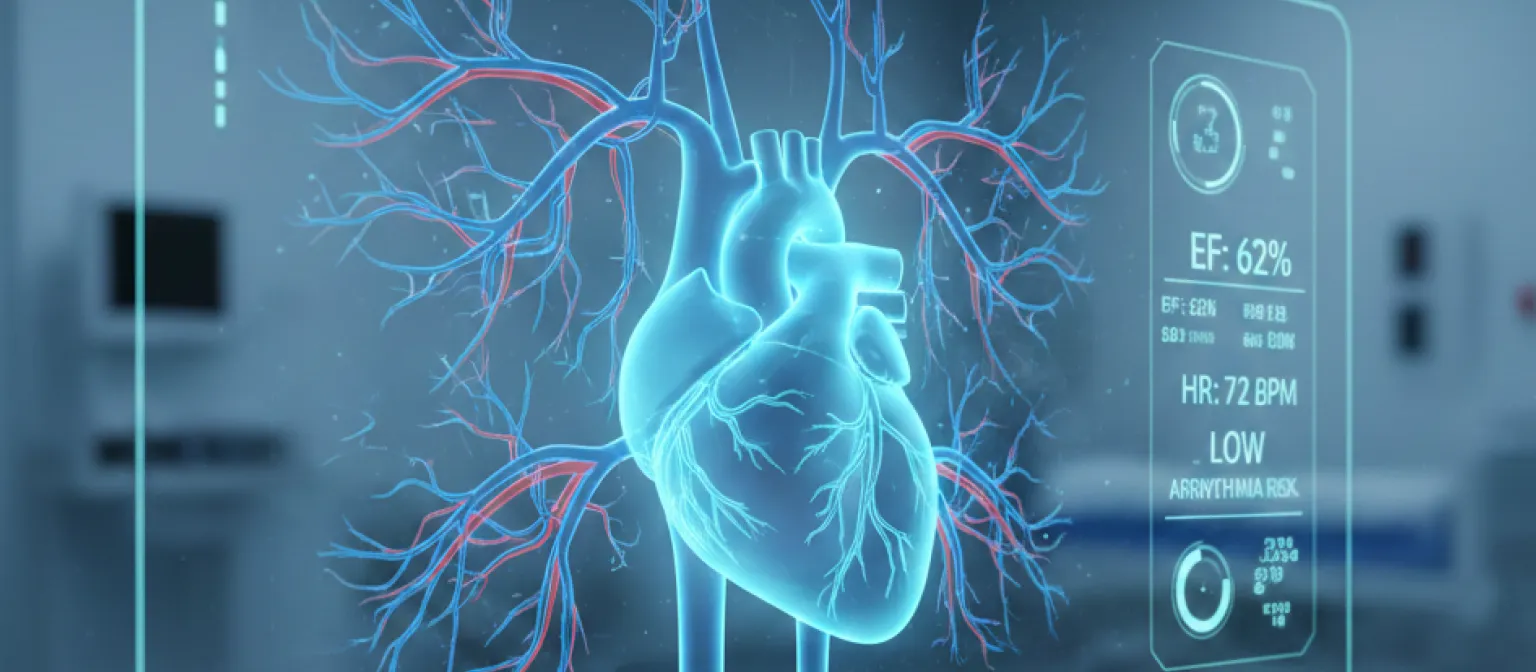
- Auto Quantify: Without delay, the software provides values of Ejection Fraction (EF) and Global Longitudinal Strain (GLS).
AI does not substitute the cardiologist but takes care of repetitive and time-consuming tasks, allowing doctors to diagnose quickly.
3. The Longitudinal Heart
Heart disease is a long-term illness. Therefore, the most advanced cardiac imaging in 2026 means reviewing a series of images over time rather than relying on a single examination.
This is where the role of the Unified CVIS becomes important. Previously, Echo reports were stored on one server, Cath Lab reports on another, and CT studies in a third.
Everrtechs NeoRad platform integrates all these systems so doctors can view a Timeline View showing:
- The ECG of the patient from 2024
- The Echo from 2025
- The CCTA from today
By having these tests visible on a single screen, clinicians can identify subtle changes in a patient’s condition that may be missed when studies are reviewed separately.
4. Structured Reporting Is the Only Way
Dictation of free text will become a thing of the past, especially as accreditation standards continue to tighten by 2026. Structured Reporting is becoming the standard.
Modern cardiac imaging software enforces a single well-defined reporting format. This guarantees that every report contains the required data points for registries such as ACC and NCDR.
Structured reporting transforms clinical reports into searchable databases, allowing healthcare providers to track outcomes and improve quality of care.
Conclusion: Software Is the New Stethoscope
The equipment used in cardiac imaging scans and probes has reached a phase of excellence and plateaued. Managing imaging data is now the next frontier.
In 2026, speed and integration will define competitive advantage for heart centers. Healthcare providers need a platform that integrates Echo, Cath, ECG, and CT into one smooth AI-powered workflow.
Key Takeaway
Is your cardiology department disorganized? Learn how Everrtechs NeoRad Cardiology PACS can bring the patient's heart at a glance through a single consolidated view.


